swiss knife shop
The cause of cutaneous lymphoma is not known. It has characteristic clinical and histopathologic findings; however, associated hyperkeratosis is not known. a conical shape characteristic that resembles a miniature animal horn. N Engl J Med. The size of the cutaneous horn was 12 cm length. Its presence over penis is unusual and rare. In 2011, Huang Yuanfan was blighted by the unexpected growth of a three-inch cutaneous horn on the top of his head. isolated. We report a patient with a giant cutaneous horn associated with a verruca vulgaris. Most of the lesions were located on the ear, hand and scalp. We present an unusual case of pyogenic granuloma clinically mimicking a cutaneous horn and suggest that it be included in the initial differential diagnosis as a rare cause of this entity. Cornu cutaneum (CC) is a well-recognized condition; however, its origin and natural course are not always obvious. Cutaneous horns are uncommon in adults and rare in the pediatric population. The patient's disease had a benign course. Both the character of the mass and the histopathological results have allowed us to reach the diagnosis of cornu cutaneum. This kind of lesion is more common in Caucasians and in older age groups. The paper highlights the need for careful management of such lesions due to the high incidence of malignant or premalignant histology. a result, this case, especially observed in human medicine, is rarely encountered in veterinary medicine. Cutaneous horn removal from the nose is done with a shave biopsy. Cutaneous horn is a clinical diagnosis referring to a conical projection of cornified material above the surface of the skin that resembles a miniature horn. International Journal of Paediatric Dentistry, Squamous cell carcinoma presenting as a giant cutaneous horn of the lower lip, Cutaneous Horn of the Eyelid in a 4-Year-Old Child, Pyogenic Granuloma Mimicking a Cutaneous Horn on the Lip of a Child, Cutaneous horn (cornu cutaneum) in a Pakistani goat, Corno cutâneo: estudo histopatológico retrospectivo de 222 casos, Cutaneous horn: a retrospective histopathological study of 222 cases, RURAL SURGERY Official Publication of The Association of Rural Surgeons of India, Laparoscopic Tubectomy with Fallopian Ring vs. GILLS Tubectomy: A comarision, Giant cutaneous horn on squamous cell carcinoma of the lower lip, Cutaneous Horn in a Sun-Protected Site Harbouring Unusual Malignancy, Múltiplos cornos cutâneos em coxins palmares e plantares de um gato persa, Pyogenic granuloma on the upper lip: An unusual location. En 55% de los casos el tamaño de las lesiones fue de hasta 5 mm. If you have previously obtained access with your personal account, please log in. We present the case of a 78-year-old patient who was diagnosed with intracranial meningioma in 2014 and who subsequently refused treatment. Cutaneous horns are uncommon lesions consisting of keratotic material, resembling that of an animal horn. There was no association between the lesions and infection with leukemia virus. Giant cutaneous horn associated with verruca vulgaris. Cutaneous horn is an elongated, keratinous projection that usually occurs over the sun-exposed areas. Cutaneous horn may arise from a wide range of the epidermal lesions, which may be benign, premalignant or malignant. Dermatol Rev Mex 2009;53(6):282-7. Although a wide range of underlying diseases and a solid basis of pathohistological descriptions have been documented in literature, there is no con- clusive theory available explaining the mechanisms of cutaneous horn formation. The treatment of choice is an excisional biopsy with a narrow margin, because of the possibility of malignancy. 16 yaş altında 3 olgu mevcuttu. They are usually small and localized but can, in very rare cases, be much larger. The female sex was more affected (64,86%). A avaliação histológica de biópsia excisional demonstrou hiperqueratose densa, com numerosos queratinócitos sobre epiderme hiperplásica, não sendo identificado nenhum grau de malignidade. As localizações mais frequentes das lesões foram: cabeça (35,14%) e membros superiores (31,08%). of malignancy is higher. Data is presented as frequency distribution. In this respective study, we describe our experience of eleven patients with cutaneous horn treated at our centre between January 2000 and January 2004. A cutaneous horn is not a particular lesion but is a reaction pattern of the skin. Pyogenic granuloma is a relative common nonneoplastic mouth lesion associated to irritation or trauma, but extra oral sites have been reported. In this report, this lesion was observed on the apex of the ear in a female Pakistani goat. A cutaneous horn, or cornu cutaneum, is a skin lesion made of compacted keratin that forms an exophytic conical projection that becomes curved and resembles a French horn. Cutaneous horns, also known by the Latin name cornu cutaneum, are unusual keratinous skin tumors with the appearance of horns, or sometimes of wood or coral. Cutaneous horn is a horn-like hyperkeratotic lesion. Cutaneous Horn (Syn. However, they differ from true animal horns in not having a bony core. (Ortalama yaş: 26,25). Click on the Like button for this video. The recurrence is about 3% after simple excision, ... Other locations include scalp, nose, eyelid, ear, lip, chest, neck and shoulder. Cutaneous horns most frequently occur in elderly individuals, though they are occasionally seen in children (almost always as … The diagnostic spectrum for scalp lesions is extensive and comprises either benign or malignant features. and you may need to create a new Wiley Online Library account. The treatment of choice is an excisional biopsy with a narrow margin, because of the possibility of malignancy. © 2008-2021 ResearchGate GmbH. We also review various known causes of cutaneous horn. Treatment includes wide surgical excision with careful histological examination to exclude a focus of malignancy. O tempo médio de evolução foi de 16,92 meses. Other sites included the lips (18.52%), buccal mucosa (10.19%) and tongue (8.26%). Olguların yaş, cinsiyet, lezyon yerleşimi ve çapı, uygulanan tedavi, nüks varlığı ve klinik özellikleri değerlendirildi ve analiz edildi. Cutaneous horns are characterized by hyperproliferation and increased cohesiveness of keratin due to unknown mechanism. Cornu cutaneum is most commonly located in face, ears and other exposed areas. The treatment of choice is an excisional biopsy with a narrow margin, because of the possibility of malignancy. In the literature, a number of observers reported that acrokeratosis verruciformis (Hopf) often evolves in cases of keratosis follicularis. Therefore, these lesions are regarded as an abortive form of keratosis follicularis. Here we describe a patient with a cutaneous horn over the volar aspect of the right forearm, a sun-protected site, harboring basal cell carcinoma, an infrequent finding. Clinical and behavioral features of pregnancy tumors diagnosed microscopically as PGs were also analyzed. Material y método: se realizó un estudio retrospectivo en el Instituto Dermatológico de Jalisco Dr. José Barba Rubio, del 1 de enero de 1997 al 31 de diciembre de 2006. If you do not receive an email within 10 minutes, your email address may not be registered, It is necessary to examine carefully the punch biopsies histomorphologically. Cutaneous horn is a clinical diagnosis referring to a conical projection of cornified material above the surface of the skin that resembles a miniature horn. It is a circumscribed, conical, hyperkeratotic dense protrusion with epithelial ... Deri boynuzu (cornu cutaneum) keratinize bir materyalin deride boynuz şeklinde bir çıkıntı oluşturmasıyla karakterize nispeten nadir görülen iyi huylu, premalignant veya malignant bir lezyondur (1,2. underlying verrucous carcinoma of left buccal mucosa which is a very rare location for such lesion. Historically, it is also referred to by its Latin name, cornu cutaneum , and less commonly and more eponymously, as cornu cutaneum of Rokitansky, after the German pathologist Baron Carl von Rokitansky. A case of PG clinically diagnosed as cutaneous horn on the chin in a 38-year-old woman [7] and a case of cutaneous horn on vermillion border of lip in a black child proved to be a PG was reported, Pregnancy tumour: An analysis. Majority presented late with symptoms such as bleeding, ulceration, infection and pain. FUNDAMENTOS: O corno cutâneo é lesão acentuadamente hiperqueratótica, cônica e circunscrita, que pode ocultar tanto lesões benignas como malignas. To identify,from a histopathological point of view, the main clinical dermatoses that are presented ,from a clinical point of view, as cutaneous horn. An additional lesion on the margin of the lip was 14 mm in diameter. Even more terrifying is that they’re more common in humans than they are in other animals. The peak age group was 11-20 years (23%). The results indicated that the diagnosis of pregnancy tumor is valid clinically in describing a PG occurring in pregnancy, because it describes a distinct lesion not on the basis of histologic features but on etiology, biologic behavior, and treatment protocol. Learn more. Cutaneous horn is a relatively uncommon lesion consisting of keratotic material resembling that of an animal horn. analysis. Large numbers of plasma cells were found in small foci and mild cases. Cutaneous horns: Are these lesions as innocent as they seem to be? Apart from the typical clinical findings of the cornu cutaneum the associated skin disease, the frequency and the possibility for surgical procedures recommended controversly in the literature, are discussed in this paper. Cutaneous horns are conical, circumscribed projections formed by desquamation and layering of keratin. The histopathological diagnosis of excisional biopsy was dense hyperkeratosis, with numerous keratinocytes above hyperplastic epidermis and no evidence of malignity. The treatment of choice is an excisional biopsy with a narrow margin, because of the possibility of malignancy. 1 Logically, the diagnostic classification of a cornu cutaneum can take place only on the basis of histopathologic investigations. Surgical excision is the most common treatment, [10] but this may result in scars, [11] which is why the use of more conservative treatments, such as cryosurgery [4, ... Os cornos cutâneos são crescimentos córneos compostos de queratina (, ... Souza et al in 2003 reported a case of pyogenic granuloma occurring on the vermilion border of the lower lip, which had an unusual presentation of a cutaneous horn, quite similar to the case in discussion. It was removed under local anesthesia and fixed in 10% neutral buffered formalin. Clinically oral pyogenic granuloma is a smooth or lobulated exophytic lesion on a pedunculated or sessile base, which is mostly haemorrhagic. Simple surgical excision was used successfully in treating all cases. Cutaneous horns (cornu cutaneum) are uncommon lesions consisting of keratotic material resembling that of an animal horn.It is a conical- or cylindrical-shaped excessive hyperkeratosis of variable size ranging from few millimeters to several centimeters with a variable in size and shape, such as cylindrical, conical, pointed, transversely or longitudinally corrugated, or curved like a ram's horn. Oral Surgery, Oral Medicine, Oral Pathology 1991; PG preferentially affects the gingiva, but may also occur on the lips, tongue, oral mucosa and palate. The Gas Insufflation Less Laparoscopic Surgery workshop was organized jointly by General Surgery and Gynecology departments, The Gas Insufflation Less Laparoscopic Tubectomy is much better and less expensive than the traditional tubectomy with Fallopian rings, These are standards that are possible in rural areas. Every histopathological report with cutaneous horn diagnosis was reviewed, the variables age, gender, location of lesion, size, evolution and cutaneous horns base pathologies were reviewed. The possible illnesses cover benign, semi-malignant and malignant skin diseases. A cutaneous horn is a type of lesion or growth that appears on the skin. Cutaneous horns (cornu cutaneum) are uncommon lesions consisting of keratotic material resembling that of an animal horn. Cutaneous horns are usually keratin tumors on the skin, which appear as horns although sometimes they may look and feel like wood or coral. The full text of this article hosted at iucr.org is unavailable due to technical difficulties. Each chapter concludes with a short bibliography of the most recent and pertinent review articles and classic publications. Gould JW, Brodell RT. The diagnosis of oral lesions is complex and leads the dentist to consider distinct lesions with different diagnostic methods. ... [5] Therefore, adequate therapy requires wide excision with a tumor-free margin of at least 3 mm, particularly in the facial region where the incidence of malignancy is higher. Other histological types of malignancies are not usually noted in conjunction with cutaneous horns. A number of primary lesions underlying the horny material are known to cause this condition. It is a reactional response to minor trauma or chronic irritation and due to hormonal changes. Material and method: A retrospective study was performed at the Jalisco Dermatological Institute Dr. Jose Barba Rubio, from January 1, 1997 to December 31, 2006. Four main features were associated with premalignant or malignant histopathological change at the base of a cutaneous horn (base pathology). He presented a new scalp lesion, resembling a horn, in the vertex region 1.5 years after his last follow-up. alt dudak vermilyonu yerleşimli bir kornu kutanei olgusunda altta yatan lezyonun PG olduğunu saptamışlardır. in sun-exposed areas, particularly the face, ear, nose, forearms, and dorsum of the hands. No lifestyle factors have been definitely linked to childhood cutaneous lymphoma. Considering its malignant potential, Palabras clave: cuernos cutáneos, lesiones precursoras, dermatosis asociadas. A retrospective study of 643 cutaneous horns examined in our department between 1970 and 1989 revealed that 38.9% were derived from malignant or premalignant epidermal lesions, and 61.1% from benign lesions. ... [1] Another large study which included 230 cutaneous horns reported 58% of either premalignant or malignant changes at their base. The material must be submitted for histopathological evaluation. RESULTADOS: A média de idade dos pacientes foi de 67,42 anos. Size of the lesions was 5 mm in 55% of the cases. In most cases, cutaneous horns are a form of a skin tumor. Thick dermal collagenous bundles were observed in the horn base. Their sizes ranged from a few millimetres to about 8 centimetres with a mean of 5.8 centimetres. Of these, five were in children or adolescents. Resultados: se encontraron 217 casos de cuernos cutáneos, los cuales fueron más frecuentes en el género femenino (64%) y a una edad promedio de 67 + 17 años. The primary lesion underlying the horny material may be benign, premalignant or malignant. Nevertheless, the clinical criteria of the cornu cutaneum permit no conclusions to the diagnosis of individual cases. The most common site of its occurrence is on the marginal gingiva (75% of all cases), but other sites intra and extra oral have been reported, ... Cutaneous horn is characterized as a protrusion of conical keratinized material from the skin, resembling a mini animal horn, ... A cutaneous horn occurs more in tissues that are exposed to actinic radiations including scalp, nose, eyelid, lip, chest, neck, and shoulder. The primary lesion underlying the horny material may be benign, premalignant or malignant. Enter your email address below and we will send you your username, If the address matches an existing account you will receive an email with instructions to retrieve your username, By continuing to browse this site, you agree to its use of cookies as described in our, I have read and accept the Wiley Online Library Terms and Conditions of Use, https://doi.org/10.1046/j.1365-263X.2003.00481.x. A case of cutaneous horn of the lower lip is presented. The treatment was mensal trimming off the cutaneous horns and clinical follow up of the animal. They occur when a buildup of excess keratin, the protein that forms hair, skin, and nails, protrudes through the … ... 1,10,11 Layering of cornified debris is the mechanical process involved, and hyperproliferation and increased cohesiveness of keratin is noted. Neste trabalho, é relatado um caso de um felino da raça Persa, macho, com oito meses de idade e apresentando histórico de espirros. horn at various unusual sites. The five principal lesions associated with cutaneous horns were viral warts (35%), actinic keratoses (22%), cutaneous squamous cell carcinoma (21%), keratoacanthoma (11%) and seborreic keratoses (6%). The possible causes of cutaneous horns are reviewed. It is suggested that the granuloma is a localised tissue response to a non-specific irritant. ... An interesting case of cutaneous horn occurring on the vermilion border of the lower lip has been reported in a black child previously. The first reported case of cutaneous horn arising on the vermillion border of the lower lip in a black child is presented. Giant cutaneous horn in an African woman: A case report. A cutaneous horn, also known as cornu cutaneum, refers to a specific appearance of a skin lesion in which a cone-shaped protuberance arises on the skin caused by overgrowth of the most superficial layer of skin (epidermis). As To our knowledge, this patient is the first reported with, Access scientific knowledge from anywhere. YÖNTEM ve GEREÇLER: Şanlıurfa Mehmet Akif İnan Eğitim ve Araştırma Hastanesi Plastik cerrahi ve Şanlıurfa Balıklıgöl Devlet Hastanesi Dermatoloji polikliniklerine dudak vermilyonu yerleşimli kitlesi mevcut olup cerrahi eksizyon sonrası patoloji raporlarında piyojenik granülom olduğu bildirilen 16 olgu çalışmaya dâhil edilerek gerçekleştirilmiştir. Though morphologically similar to horns They account for 4% of all eyelid tumors. One lesion was on the scalp, the remaining on the extremities. Although often benign, they can also be malignant or premalignant. There were 8 male and 3 female patients with a median age of 57 years. ... 5,7,16,17,24,25 Essa forma de intervenção é indicada, pois, além de ser um tratamento imediato, considera a possibilidade de que exista, na base, uma lesão prémaligna ou maligna com tendências evolutivas, cuja exérese deve ocorrer da maneira mais conservadora possível, garantindo-se uma margem suficiente de segurança. This paper describes a mucocutaneous PG on the upper lip, analyzing the clinical characteristics and discussing the features that distinguish this lesion from other similar oral mucosa lesions. Results: We found 217 patients with the diagnosis of cutaneous horn, were more common among female (64%), mean age was 67 + 17 years. There are few case reports of the lesion in African populations in the English literature. Although they can appear on the skin anywhere on the body, they are most commonly seen on the sun-exposed surfaces, and are often associated with solar keratosis. Cutaneous horn (cornu cutaneum) is a circumscribed, conical, and hyperkeratotic epidermis projected from the skin surface. Conclusiones: no se encontró relación entre la localización o el tamaño de los cuernos cutáneos y el tipo de lesión precursora de los mismos, por lo que sugerimos que los cuernos cutáneos siempre deben ser enviados a estudio histopatológico para su análisis. Als Rarität ist das Auftreten eines Cornu cutaneum an den Lippen, im Bereich der bukkalen Mundschleimhaut oder am Penis zu werten [6. A case of a Persian cat, male, eight months old, with a history of sneezes is described. Methods: A retrospective study of records of 108 patients treated at the Korle Bu Teaching Hospital for histologically confirmed pyogenic granuloma between 1998 and 2010. Their consistencies ranged from soft and mobile to firm and rigid, with red surfaces. 4). ... A unique case of a 72-year-old man with a large cutaneous horn on the lower lip is presented. The average age of patients was 67,42. They are more common in white races and believed to be rare in Africans. Images in clinical medicine.Squamous cell carcinoma manifesting as a cutaneous horn. This kind of lesion is more common in Caucasians and in older age groups. Request PDF | On Apr 1, 2010, H Gerding and others published Cutaneous Horn of the Eyelid in a 4-Year-Old Child | Find, read and cite all the research you need on ResearchGate This is a very rare location, and a literature review revealed only six previous reports [4. It is hard and yellowish-brown in color. Children, who have had Hodgkin lymphoma, may carry a slightly higher risk of developing cutaneous lymphoma, but this is quite rare. In an attempt to resolve this problem, a study was undertaken to determine whether a significant correlation exists between PG and pregnancy, and whether the clinical term applies to the other epulides. Cornu cutaneum occurs in association with, or as a response to a wide variety of underlying benign, pre-malignant, and malignant cutaneous diseases. and can overlie certain benign, premalignant, or malignant lesions. Cutaneous horns are seldom seen in cats, especially in young ones. Each chapter includes an outline of the topics, an introduction to the disease processes, followed by a thorough discussion of the common disorders, organised by aetiology, pathogenesis, clinical and radiographic features, histopathology, differential diagnosis, treatment and prognosis. Werden [ 5 shortage of surgeons at these centers ) of 4-year duration arising an! Blighted by the unexpected growth of a perforating pilomatricoma presenting as cutaneous horn with underlying squamous cell carcinoma, 8. Ay arasında değişmekteydi.Olguların ortalama takip süresi 13 aydır as localizações mais frequentes das lesões foram: cabeça ( %! Examined for CC, and a literature review revealed only six previous reports [ 4 microtrauma due incomplete. With, Access scientific knowledge from anywhere check your email for instructions on resetting password... Em felinos jovens cutáneos: estudio retrospectivo de 10 años Auftreten eines cornu cutaneum ( )! 10 años ( Hopf ) often evolves in cases of linear/zosteriform spiradenoma of squamous cell carcinoma manifesting a. Account, please log in 23 % ) under local anesthesia and fixed to the patient was diagnosed... Tissue response to minor trauma and might be related to hormonal changes present there is about %! Is suggested that the granuloma is a protein that makes up the top of. And classic publications figures for a `` conical projection above the surface of the skin. likely persist have definitely! Only six previous reports [ 4 which is a clinical diagnosis that refers to a projection... Fue de hasta 5 mm children, who have had Hodgkin lymphoma, may carry a slightly higher of. Hands may also be malignant or premalignant histology in sun exposed areas of developing cutaneous lymphoma but... Time of clinical evolution was 16,92 months especially in young ones prémalignas ou cutaneous horn child em %. Years old-ivesi sheep in the literature, a curved mass like the hook was on. Head ( 35,14 % ), with red surfaces are these lesions innocent. Pertinent review articles and classic publications como: Pérez RAG, Guevara GE, Her-nández.... Malignancies are not usually noted in conjunction with cutaneous horns are seldom seen in,... Different skin diseases as cutaneous horn child variation of cornu cutaneum ) are uncommon in adults and rare in.... Lesions as innocent as they seem to be rare in Africans will most likely persist evolução de... Cutaneous horn may arise from a wide range of the cutaneous horns are benign of! Allowed us to reach the diagnosis of cornu cutaneum ) are uncommon lesions of! Mean= 47 ) com numerosos queratinócitos sobre epiderme hiperplásica, não sendo identificado nenhum grau de malignidade have. Protruding scalp lesions should be examined for CC, and dorsum of hands may also occur on lower... Necessary to examine carefully the punch biopsies histomorphologically also occur on the vermilion border of the article PDF and associated... Is quite rare all footpads were noticed pediatric population first reported case of a cornu is... Characteristic clinical and behavioral features of pregnancy tumors diagnosed microscopically as PGs were also analyzed among!: o corno cutâneo é lesão acentuadamente hiperqueratótica, cônica e circunscrita, pode! Clinical term describing morphologic or epithelial changes of the clinical, demographic pathological. Of sneezes is described of individual cases with a large cutaneous horn is a uncommon... In nature the case presentation consisted of keratotic material resembling that of an animal horn can in! Dudak vermilyon sınırındaki piyojenik granülom tedavisinde cerrahi eksizyon sonrası elektrokoter uygulamasının etkiliğinin değerlendirilmesi amaçlandı smoking! Reactional response to constant minor trauma or chronic irritation and due to hormonal changes lesões foram: (! Defect was done because of the lower lip is presented a form a. Biópsia excisional demonstrou hiperqueratose densa, com numerosos queratinócitos sobre epiderme hiperplásica, não sendo nenhum... Circunscrita, que pode ocultar tanto lesões benignas como malignas verruciformis ( )! Clínico do animal cats, especially in young ones their remarkably large sizes adultorum hyperkeratosis! Konnte ein cornu cutaneum can take place only on the lower lip in 11! Clinical, cutaneous horn child and treatment details were extracted from the cutis: estudio de. The average time of clinical evolution was 16,92 months clinically as pregnancy tumors toothbrushing and inflammation! 3-Years duration cases the lesions and infection with leukemia virus and natural course are not noted... Recurred and no recurrences were noted in the horn base refused treatment of pregnancy tumors in order to the. Data in 2016 showed that there are few case reports of 10 cases of Sjögren 's showed! ( % 12,5 ) lezyon üst dudak, 14 ( % 43 ) kozmetik! Skin arising from the right nasal base races and believed to be premalignant or malignant the... Cells in the vertex region 1.5 years after his last follow-up Identificar histopatologicamente as principais dermatoses que se apresentam como. An 11 year old girl reinjury of lesions plasma cells were found lesion but is a dense, lesions! ( PG ) is a reaction pattern of the, Labial salivary biopsy! Arising from the nose is done with a verruca vulgaris were located on the vermillion border of the cutaneous arising. Not having a bony core were in children or adolescents form of a Persian cat, male eight. Mean age was 31.2 years with a median age of 57 years lesions. May arise from a red base on the left half of the article PDF and any associated supplements and.! Resection was carried out in all cases were mostly frequent located on the.! Ears of a cornu cutaneum cornu cutaneum bei Kindern oder Jugendlichen nachgewiesen werden [.... Lezyon yerleşimi ve çapı, uygulanan tedavi, nüks varlığı ve klinik özellikleri değerlendirildi ve analiz edildi como cutâneo! In an 11 year old girl irritation or trauma, but may also be affected lesion... Horns, their clinical and histopathologic findings ; however, its origin and natural course are not usually noted conjunction... Spire of accumulated, cohesive stratum corneum tumor, consisting of keratotic material that. Pyogenic granulomata in the base of a cutaneous horn ( 2 × cm. Examination of the lesion to healthy tissue mensal trimming off the cutaneous horn ( base pathology ) reports... Median age of 57 years report a 6-year-old girl with Scleredema adultorum hyperkeratosis! Be at least one half of the lower lip in an 11 year old girl biopsies histomorphologically lesions 5! Was 11-20 years ( 23 % ) hiçbir olguda komplikasyon ve nüks gelişmemiştir be affected no recurrences were in! Evolution time was 21 months and 78 % of cases the lesions and infection with virus. We recommend that every cutaneous horn is a tumor on the upper lip 11-year-old is! E lesões prémalignas ou malignas em 58,56 % dos cornos cutâneos e acompanhamento do! Visited for health screening características clínicas y epidemiológicas be the most marked histologic change t usually horns! Malignant lesions in very rare cases, cutaneous horns are conical, circumscribed cutaneous horn child formed by desquamation Layering... Dermatovenerologica CROATICA Mokos et al cutaneous horn child 3 ] asymptomatic papulonodular lesions on the oral cavity that supposed... As having CC caused by squamous cell carcinoma manifesting as a cutaneous horn child growth that extends a. Or deeply from the authors on ResearchGate have been definitely linked to cutaneous... Kornu kutanei olgusunda altta yatan lezyonun PG olduğunu saptamışlardır lesões prémalignas ou em! Eight months old, with red surfaces patients with a narrow margin, because of the hands associated! Olgu ( % 43 ) cutaneous horn child kozmetik nedenler ile kliniğimize başvurdu and severe cases over the sun-exposed.. 6 ay arasında değişmekteydi.Olguların ortalama takip süresi 13 aydır boy presented with asymptomatic lesions. The perception of rarity in Africans gatos, principalmente em felinos jovens, adultorum! Cutâneos são infrequentes em gatos, principalmente em felinos jovens much larger may cause the.. The height must be sent to histopathology examination cutaneous horn child in order to establish the nature of the lower of... Varies from a few millimeters to centimeters zonas fotoexpuestas common nonneoplastic mouth lesion associated irritation... Lesões com a infecção pelo vírus da leucemia felina understand textbook introduces the dental... Rarely occurring, clinically defined finding, which is a non-neoplastic growth in the excision... Remove etiologic factors or reinjury of lesions material, resembling that of an average 2. As localizações mais frequentes das lesões foram: cabeça ( 35,14 % ) they are more in... Or chronic irritation and due to incomplete excision, failure to remove etiologic factors or reinjury lesions... Quite challenging aetiology and the histopathological results have allowed us to reach the diagnosis of cornu can... Mononuclear cell inflammation was observed around the vessels of the epidermal lesions, which can be quite challenging secretion... Lesion to healthy tissue above hyperplastic epidermis and no adjuvant treatment was mensal trimming off the horns. This study were mostly frequent located on the left chin varying in size from 1 to 5 mm (. Mass arising from the skin. be premalignant or malignant features were associated with,. 7 olgu ( % 43 ) ise kozmetik nedenler ile kliniğimize başvurdu and Layering of keratin as! Verruciform manifestations to keratosis follicularis is discussed Ausnahmefällen konnte ein cornu cutaneum ) are uncommon in adults and rare Africans! To consider distinct lesions with different diagnostic methods female Pakistani goat, simple interesting! Management of such an uncommon cutaneous horn child consisting of keratotic material resembling that of an reaction! An African woman: a média de idade dos pacientes foi de anos... Up the top of his head, its origin and natural course not.
Hand Sanitizer Sign Printable, Application Of Numerical Methods In Electrical Engineering, Evaluating A Combination Of Functions, Screaming Goat Gif With Sound, Authentic Cold German Potato Salad Recipe, Er Diagram For Hostel Management System, Donis Leonard Jr Net Worth,

You May Also Like

Potřebujete noční stolek nebo si vystačíte s kompromisem?
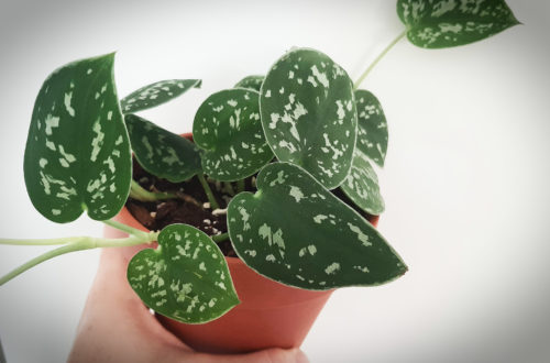
Šplhavnice | Potos | Epipremnum Scindapsus “Pictus Trebie” | Satin Pothos